 |
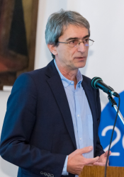
| Mihály Pósfai at the briefing |
The successful application for funding was partly due to the high-quality research activities of his research group. In 2015 the professor was awarded a discovery research grant in the framework of the Researchers' Thematic Applications Programme.
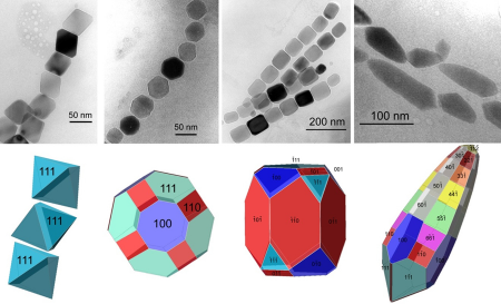
“Indeed, these projects are related. I am a geologist by profession; I have always worked with minerals and nano-objects from the very beginning. You could say that it was by sheer coincidence that I started to study bacteria which develop magnetic nanocrystals in human cells. This makes them behave like compasses: they have to swim were the Earth's magnetic field directs them. These organisms are not only exciting for biology but also for materials science as they develop magnets with regulated size and shape – and this is something we can hardly do even in laboratories. In one of our funded research projects the regulation mechanism identified in a magnetic bacterium is applied under laboratory conditions to create one-dimensional magnetic structures.”
 |
| Magnetic bacterial cells containing magnetic nanoparticles in various shapes and array |
Will there be any practical use of these achievements?
“One of my PhD students has already created such structures and it turned out that – compared to “traditional” dispersed magnetic nanoparticles – they increase the viscosity of certain liquids by several orders of magnitude. In fact, any magnetic material with a high level of anisotropy (i.e. has a special elongated shape) is a potential field of application. One-dimensional formations can be useful for example in data storing or magnetic resonance imaging (MRI).
The other project focuses on how carbonate minerals are formed in Lake Balaton. The Lake’s mud is largely made of magnesium calcite which precipitates in the water. I have said that the two research projects are related because micro-organisms also play a part in mineral precipitation. The photosynthesis performed by algae creates suitable chemical conditions for the precipitation of magnesium calcite. Calcite does not only appear “in itself” in the solution, by the formation of homogeneous crystal germs, but it precipitates on a surface. Lake Balaton is shallow and its water is always full of tiny floating clay mineral particles providing a surface for calcite to crystallise. In winter, when the water freezes over, crystallisation sets in on the cells.
In the case of magnetic nanofibers the inorganic material is crystallised to mutagenic filaments to control its precipitation. In Lake Balaton natural control is ensured by clay minerals or suitable microalgae. Studying this process is interesting to science because it seems that apart from the long-established classical crystal germ formation mechanism; there are other processes in action as well. The new election microscopy laboratory will be a great help in monitoring them.”
What are the main elements of the project aimed at establishing the electron microscopy laboratory?
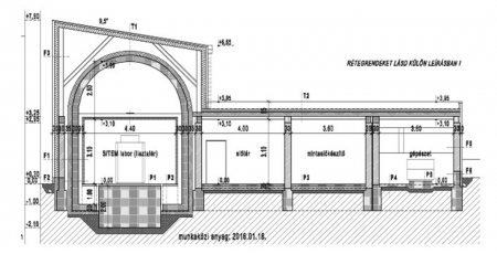
“The funding will be spent on two microscopes: the old scanning electron microscope will be replaced with a new one, and a transmission electron microscope will be installed in a new separate building. The new building indicated in our application is very important as the microscope located here can only be used in a noise-free environment.”
What does noise mean in this context?
“Partly mechanical noise, vibration: the “standard vibration” of the city reaches the microscope through the rocks and the soil which frustrates the atomic resolution, the detection of individual atoms. These microscopes will not be installed in the first floor but in the basement, and even the environment of the new building will be adapted to the device. Electromagnetic noise will also have to be filtered: for example, we have to pay attention to what kind of devices are operating in the building and how wires run in the wall. Acoustic noises also cause disturbances but these modern microscopes mostly come in big boxes rather than on their own and even the operator controls the test from another room.
As I have referred to it, the electron microscope makes it possible to observe solid material in nanometre sizes or even “atom by atom”. But such equipment are not only suitable for imaging but their accessories can be used for determining the composition of the examined material, and even 3D morphology and composition can be determined by "tilting” the sample (the 3D process is called electron tomography). Even biological samples can be studied but this requires special preparation. Part of the funding will be spent on two pieces of sample preparation equipment: an ultramicrotome for creating thin biological sections and an ion-beam thinner for the examination of “hard” samples.”
In your presentation you mentioned that transmission electron microscopy may mark a great leap forward in the exploration of protein structures.
“The epoch-making change is the result of a new sample preparation method: the biological sample is immersed in liquid ethane and the sudden cooling (ethane melts at -182 °C) causes the water in the sample to solidify in an amorphous form rather than in a crystallised form which prevents cell organoids from changing. The accessories required for this are not included in the present funded project, but they are included in our EDIOP project proposals pending decision.”
You are in the middle of the preparatory phase of the project. Were the first steps successful?
“We have completed an important work phase: the drafting of the public procurement notice in relation to the assets to be purchased. We now have the building permit drawings and we have developed the operating plan of the laboratory.
 |
| Excerpt of the blueprint of the laboratory under construction |
What makes the laboratory so special?
“The scanning and transmission devices will enhance the competitiveness of Hungarian researchers in this field bringing them up to par with international research groups using cutting-edge technologies.
There are many research teams at the University of Pannonia who would have needed electron microscopy for their work, and there are many others who actually use electron microscopy by bringing their samples into another facility and paying for the examination – but work is ineffective this way.
We want to create a laboratory which provides quality services to everyone, not only to the university but to a wider range of stakeholders. Institutions in our region include the University of West Hungary and the Széchenyi István University, a little further south the University of Pécs and the Balaton Limnological Institute, but we also expect industrial orders. So, we think that the laboratory will be suitable for the examination of various samples and scientific issues, and we want to operate it as efficiently as possible. Similar laboratories abroad not only give home to local staff but also to “outsiders” because the electron microscopy is very time consuming.
Our plan is that research groups wishing to use the equipment more frequently would delegate a colleague who would be trained by us to perform certain techniques and tasks. He or she would then be qualified to carry out such tasks and would be allowed to use the microscope independently. It is always more effective to allow researchers bringing the scientific issue to carry out the measurements as they the most familiar with the given problems. Naturally, we undertake to answer simple questions and we have our own research topics as well.”
Would you use this revenue to maintain the laboratory?
“We expect the laboratory to be self-sustaining: the revenue would be spent on staff wages, material costs, microscope maintenance costs, and we would also form a reserve for depreciation to cover the costs of future developments on the laboratory.”
---------------------------------------------------------
Funded project:
Establishment of an Electron Microscopy Laboratory at the University of Pannonia (SEM: scanning electron microscope, TEM: transmission electron microscope)
Project manager: Bálint Morvai
Technical leader: Mihály Pósfai
Budget: HUF 924 million
Duration: 01/06/2016–31/05/2019
- 31/05/2017 Completion of the project preparation phase, completion of public procurement procedures, selection of the contractor
- 30/05/2018 Testing of the new SEM, delivery of the new building, deployment of the new TEM
- 31/05/2019 Launch of the laboratory, development of international R&D service functions


